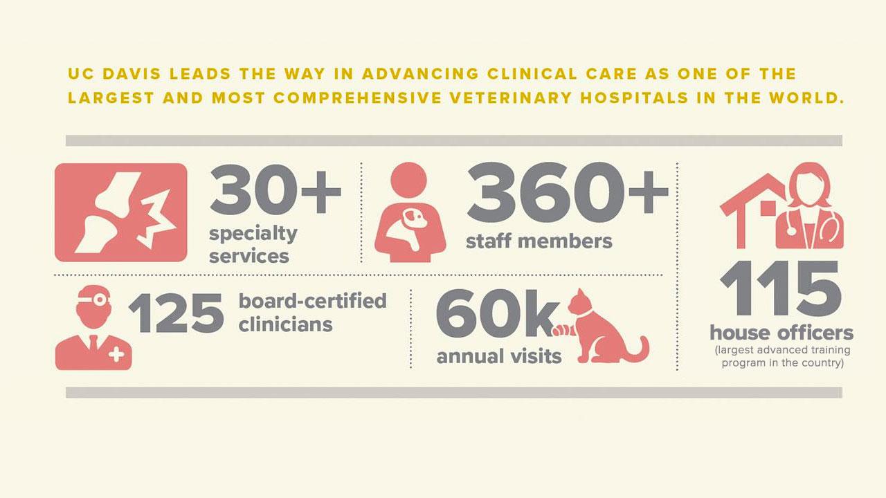
UC Davis leads the way in advancing clinical care as one of the largest and most comprehensive veterinary hospitals in the world.
As the school boldly looks to the future with its Veterinary Medical Center project, UC Davis will construct an entirely new hospital, expanding the size and scope of its current Large and Small Animal Clinics. This will allow clinicians to expand their cutting-edge procedures and continue to push the limits of veterinary medicine. With the help of benefactors and dedicated clients, UC Davis veterinarians will be able to realize their visions, as they integrate teaching, research, and clinical activities into compassionate care that will transform lives.
Some of the latest clinical advances include:
PET Scanning
Faculty Lead: Dr. Mathieu Spriet,
Diagnostic Imaging Service
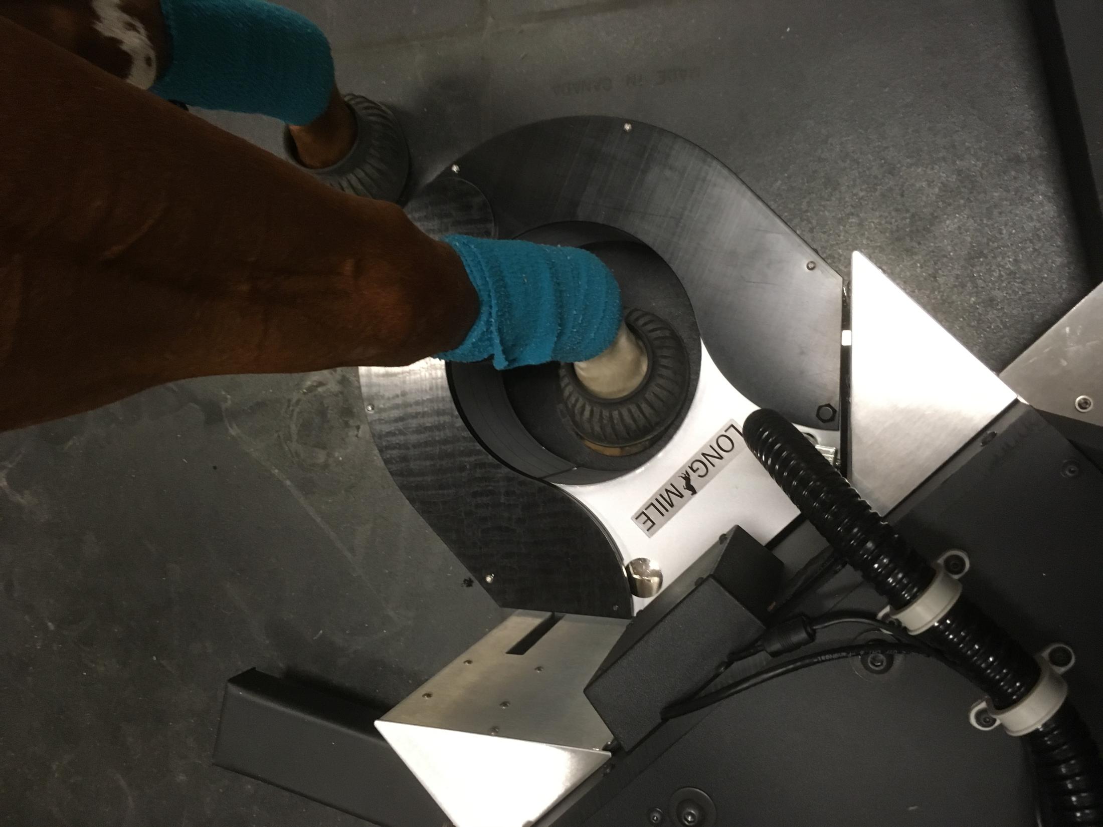
Horses requiring positron emission tomography (PET) to distinguish between active and inactive injuries in leg bone and soft tissue can undergo scanning more safely. This reduces risk inherent in general anesthesia as it only requires sedation.
Learn More
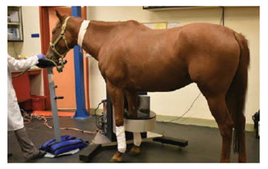
Equine PET Scanning Technology Continues to Advance Dr. Mathieu Spriet, associate professor of veterinary radiology, is advancing his positron emission tomography (PET) research and clinical translation to include a newly-developed standing PET scanner. Instead of being under general anesthesia—which is required to perform a traditional scan—a horse only needs to be sedated to perform a standing PET scan. The ability to utilize this technology on a standing horse under sedation instead of anesthesia will greatly expand the availability of this powerful imaging technique, allow for more routine use, and open it up to patients that are not able to undergo anesthesia. Dr. Mathieu SprietA recently completed phase of the study consisted of validating the safety of the system and establishing scanning protocols using research horses from the UC Davis Center for Equine Health. Six horses were imaged twice with the standing scanner and once under general anesthesia. This allowed the researchers to confirm the repeatability of findings and to compare with results obtained with the technique previously developed on anesthetized horses.
The horses tolerated all of the procedures well. All imaging sessions were successful, and no complications were reported. The quality of images obtained on the standing horses were similar to the ones performed under general anesthesia.
“We are excited to report that everything worked according to plan, if not better,” said Dr. Spriet. “I am very impressed with the quality of images we were able to obtain.”
Data used for both research and clinical applications has shown PET to be particularly useful for detecting bone lesions that are not recognized using other imaging techniques, and to distinguish between active and inactive injuries in bone or soft tissue.
Thanks to the prolific amount of research Dr. Spriet has performed since 2016 (scanning more than 100 horses), Santa Anita Park installed a standing PET scanner for early detection of pre-existing bone changes before they lead to catastrophic injuries in racehorses.
“PET has a very interesting role to play in racehorses, as it detects changes at the molecular level, before structural changes occur,” explained Spriet. “In other words, PET provides warning signs that injuries might happen. There is still a lot of work ahead of us, as we need to learn to distinguish the PET changes that reflect normal adaptation to speed work from changes that are indicative of high risk for major injuries.”
Videoendoscopy to Diagnose Upper Respiratory Disorders
Faculty Lead: Dr. Scott Katzman,
Equine Lameness and Surgery Service
Overground videoendoscopy enables veterinarians to exercise the horse in its normal environment, such as a track or an arena rather than on a treadmill, and diagnose causes for upper respiratory collapse not observed during resting endoscopy.
Learn More
Dr. Scott Katzman, chief of the Equine Lameness and Surgery Service, is utilizing overground videoendoscopy to diagnose dynamic upper respiratory collapse in horses. A malleable endoscope is inserted into the horse’s nasal passage and advanced into the nasopharynx to observe laryngeal movements during ridden exercise. The examination is recorded and transmitted to a small monitor for viewing in real time. With this technology, veterinarians can diagnose causes for upper respiratory collapse not observed during resting endoscopy.
Utilizing this technology enables the veterinarians to exercise the horse in its normal environment, such as a track or an arena rather than on a treadmill, which can be dangerous and frightening for inexperienced horses. Images obtained from the videoendoscopy greatly increases the likelihood of making a definitive diagnosis in horses presented for upper airway noise with or without associated exercise intolerance.
“When they’re not in their normal environment, not in their normal tack, and not being ridden as they normally would be, are you getting the full picture of their true condition?” questioned Dr. Katzman, explaining the benefits of the system. “Viewing them while they’re actually exercising normally, we are able to get a more genuine view of their upper respiratory tract in action.”
Stem Cell Therapy for Equine Athletes
Faculty Lead: Dr. Larry Galuppo,
Equine Lameness and Surgery Service
Stem cell therapies offer promise due to these cells’ ability to replicate, regenerate tissue, and repair damaged tissue.
Learn More
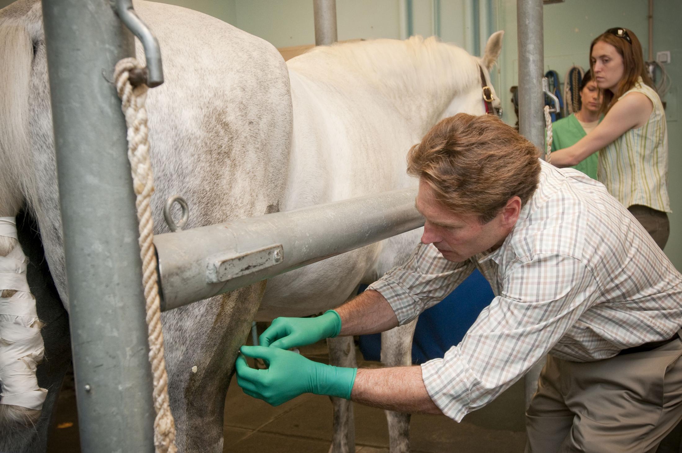
Dr. Larry Galuppo administers stem cells to a horse as part of a clinical trial at the UC Davis veterinary hospital.Tendon and ligament injuries are a common cause of lameness in horses. Treatments vary greatly and are often associated with high expenses and failure to return to a previous level of performance. Non-elastic scar tissue formation is a frequent result of injury. Since scar tissue is not as functional as tendon and ligament tissue, Dr. Larry Galuppo is exploring the use of stem cells to develop an effective treatment that will improve healing and lessen the amount of scar tissue formation.
While the clinical use of stem cells is currently only utilized through clinical trials at UC Davis, the application has shown positive results for many horses. By engaging in standardized clinical trials and collaborating with human medicine teams, the goal of this work is to generate evidence based recommendations for stem cell therapies.
Dr. Galuppo is conducting four trials through the Veterinary Center for Clinical Trials. His focus is on mesenchymal stem cells’ ability to replicate themselves, regenerate tissue, and repair damaged tissue to treat tendon and ligament injuries, intra-articular lesions, and laminitis in equine athletes.
These innovative treatment options may improve a horse’s healing process during the crucial time of potential scar formation, enhancing the quality and strength of repair. If this is the case, re-injury rates should decline and return to previous level of performance should be more common in horses treated with regenerative medicine.
In 2017, racehorse Irish Streetsinger returned to racing following enrollment in Dr. Galuppo’s stem cell trial for an injury that sidelined her in 2016. By early 2018, she ran seven races with one victory at Golden Gate Fields.
Eventually, these stem cell therapies may become integrated as a routine part of regenerative medicine for sport horses as well as human athletes suffering from similar conditions.
These stem cell projects—being developed in conjunction with the school’s Veterinary Institute for Regenerative Cures—are great examples of research and clinical faculty working together to apply “benchtop to bedside” translational medicine.
Advancements in Interventional Radiology
Faculty Lead: Dr. Bill Culp,
Soft Tissue Surgery Service
Interventional radiology (IR), which incorporates “live” imaging for guiding novel procedures, is paving the way for pioneering advancements in the treatment of our companion animals. IR is routinely be utilized to treat both benign (often urinary and vascular disease) and malignant diseases, such as cancers that lack other effective treatments.
Learn More
Interventional radiology (IR), which incorporates “live” imaging for guiding novel procedures, is paving the way for pioneering advancements in the treatment of our companion animals. Dr. Bill Culp of the Soft Tissue Surgery Service routinely utilizes IR in his practice to treat both benign (often urinary and vascular disease) and malignant diseases, such as cancers that lack other effective treatments. The different applications for IR in veterinary medicine are changing the face of the available treatment options and becoming the standard of care – many of these have been pioneered at UC Davis by Dr. Culp.
“The performance of many IR procedures is still uncommon in veterinary medicine, as advanced training and equipment is required,” Dr. Culp said. “The UC Davis veterinary hospital is one of the top institutions in the world in the field of IR, and the program is continuing to grow. Currently, we offer an extensive array of treatment options, and this is increasing regularly allowing our program to grow into one of the largest and busiest in the world. As equipment continues to evolve and become more advanced, we look forward to expanding this specialty at UC Davis, as we strive to give our patients the best possible quality of life.”
Because of these exclusive offerings, clients are bringing their pets to UC Davis from far distances, including Canada, Hawaii and the East Coast.
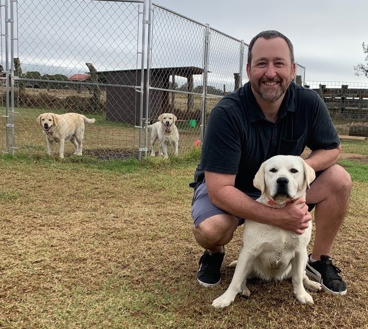
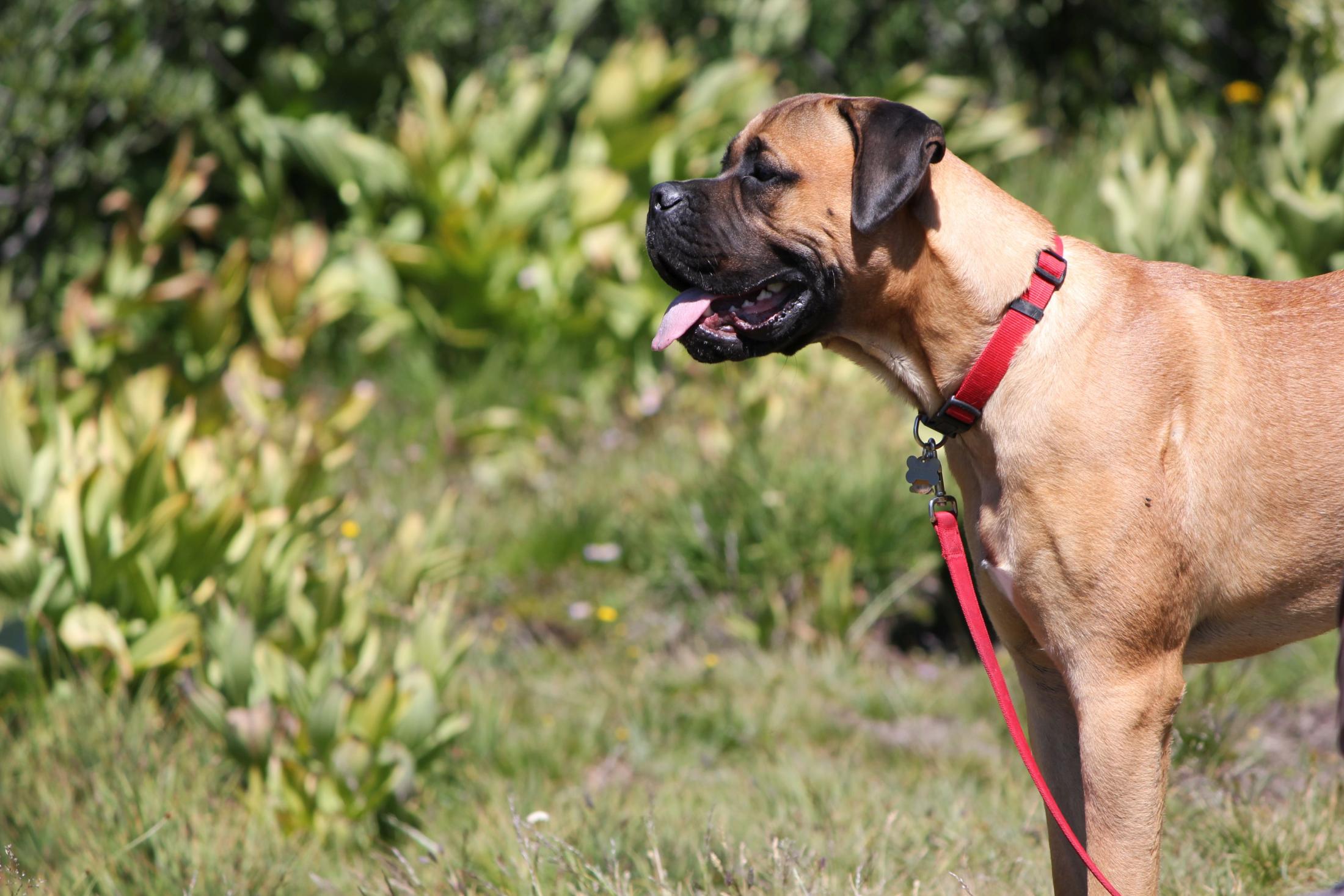
Treating Vascular Anomalies: Shunts and Arteriovenous MalformationsRhyme Rhyme, a 2-year-old male cat, was being medically managed for a liver shunt for six months. That followed nearly two years of trying to discover the root of his illness, which seemed to have plagued him for as long as could be remembered. When a specialist in Rhyme’s hometown of Vancouver, British Columbia finally diagnosed the shunt, he recommended Dr. Culp, whom he knew to be performing a minimally invasive procedure to correct shunts for many years. A liver shunt—or more specifically an intrahepatic portosystemic shunt (IHPSS)—is a birth defect of the blood vessels that should bring blood to the liver for purification. When an IHPSS exists, the liver does not receive the proper amount of blood, as the liver is being bypassed by the abnormal blood vessel or vessels. The goal of any treatment with liver shunts is to close the shunting vessel over time, thus redirecting blood flow through the liver, allowing adequate nutrition to reach the liver, as well as for toxins to be removed from the systemic circulation. Using an IR approach, Dr. Culp was able to utilize live x-ray (fluoroscopy) to guide highly specialized equipment through Rhyme’s vasculature to the shunt and perform the needed blockage. A metallic stent was placed in Rhyme’s vena cava in the region of the shunt. Metallic coils were placed into the shunting vessel to cause a reduction of blood flow through the shunt. This procedure avoided having to open Rhyme’s abdomen and made his recovery much quicker with less discomfort. Dr. Bill Culp with DJ in HawaiiDJ, a 6-month-old female yellow Labrador retriever, was brought to UC Davis by her family from Hawaii to be treated for an IHPSS. DJ’s minimally invasive procedure was performed through her jugular vein, and catheters were used to place a metallic stent in her vena cava and several metallic embolic coils in the abnormal shunting vessel. Several months later, on a vacation to Hawaii, Dr. Culp and his family randomly encountered DJ and his family, who invited them to their home to share a traditional Hawaiian meal.
After this chance encounter, Dr. Culp commented, “It was wonderful to be able to see how well DJ was doing while we were in Hawaii, not in a small part to the amazing care provided by her ‘parents,’ who demonstrated such tremendous concern for DJ’s well-being. She is now living the life that we hoped for when she underwent treatment.”
Another congenital blood vessel anomaly regularly seen in Dr. Culp’s clinical practice is called an arteriovenous malformation (AVM). Much like IHPSS, AVMs are an abnormal blood vessel communication that patients are often born with. However, with this anomaly, an artery attaches to a vein as opposed to what is seen with IHPSS where veins connect to other veins.
With AVMs, the blood supply will enter a part of the body but will generally exit that area incorrectly and without reaching its intended tissue target. In animals, AVMs are most common in the liver and distal limbs. In humans, AVMs are also regularly diagnosed in the brain. Dr. Culp is having success treating AVMs with embolization, a procedure that delivers a product to “clot” or close the blood vessels and redirect the blood flow. These procedures are performed minimally invasively, entering the body through major blood vessels like the femoral artery and navigating to the location of the blood vessel malformation.
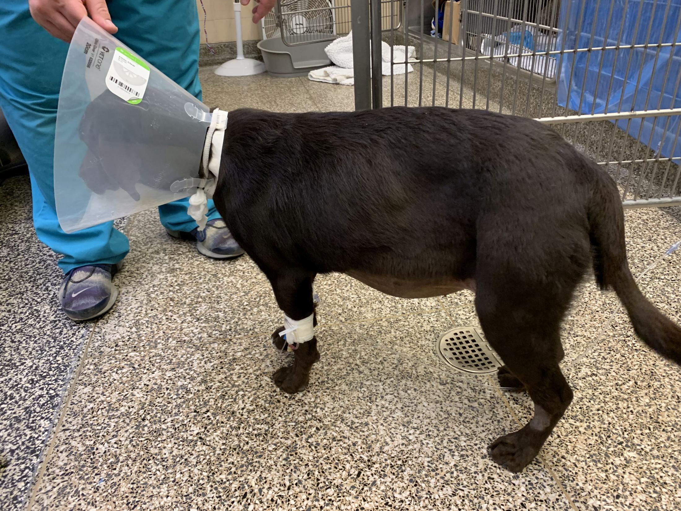
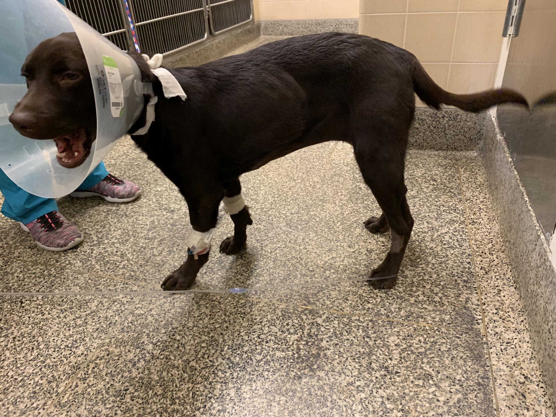
Liver AVMLuna before surgery with fluid buildup in her abdomenLuna after surgery with decrease of fluid buildup in her abdomen Luna, a 10-month-old female chocolate Labrador retriever, was diagnosed with a liver AVM by Dr. Alyse Zacuto, an internal medicine specialist in Luna’s hometown of San Diego. Dr. Zacuto is a former student and resident at UC Davis, and saw first-hand the school’s treatment for abnormal vasculature. She referred Luna to Dr. Culp, who performed a minimally invasive procedure through Luna’s femoral artery, guiding wires and catheters through this blood vessel to the AVM. The blood supply to Luna’s AVM was blocked with glue, stopping further flow through the malformation. Liver AVMs generally cause a large buildup of fluid in the abdomen. Following surgery, Luna’s abdomen showed a dramatic decrease in fluid buildup in just a few days. She continues to recover well back home under Dr. Zacuto’s care.
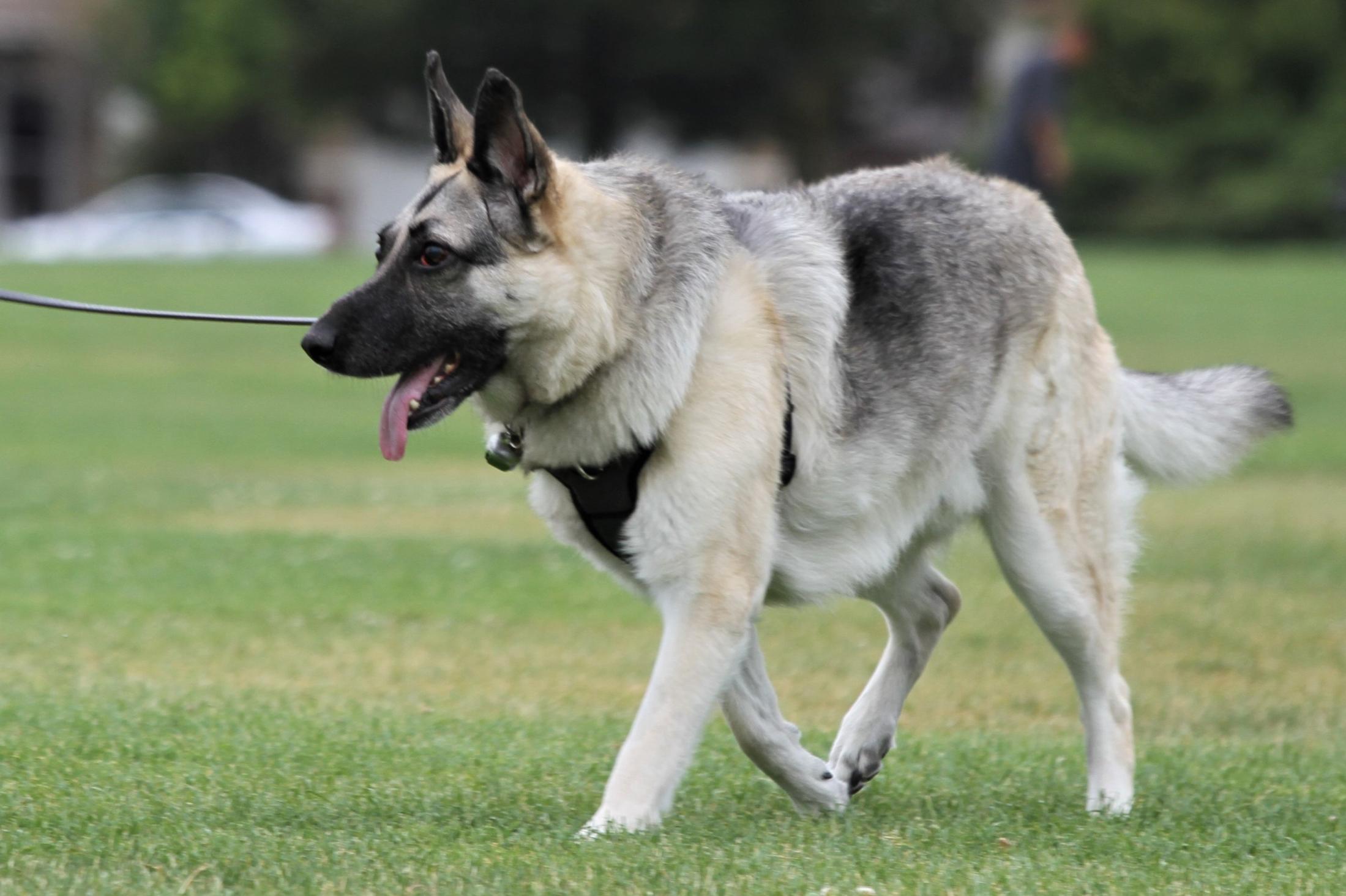
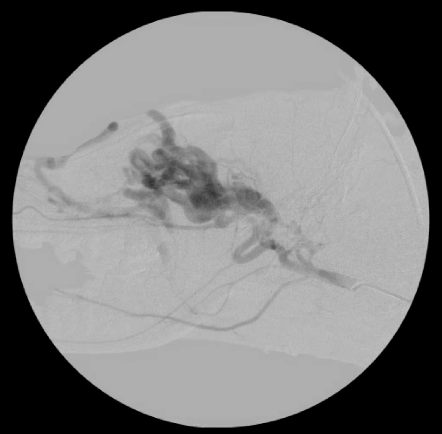
Brain AVMCrashCrash's flouroscopy showing brain AVM Crash, a 6-year-old male German shepherd, was diagnosed with a brain AVM located behind his eyes causing tremendous pressure in his head. Brain AVMs are extremely rare in veterinary patients, but the condition occurs in human patients more commonly, so Dr. Culp collaborated with physicians at UC Davis Health—an interventional radiologist and a neurointerventionalist—who were specifically trained in the treatment of vascular diseases of the brain and treat AVMs in humans on a regular basis. Together this team performed an embolization to close the blood vessels in Crash’s AVM and redirect the blood flow. Crash no longer feels the constant pain that he was experiencing in his head, and the pressure in the region of his eyes has improved.
Treating Prostate and Liver Cancer:
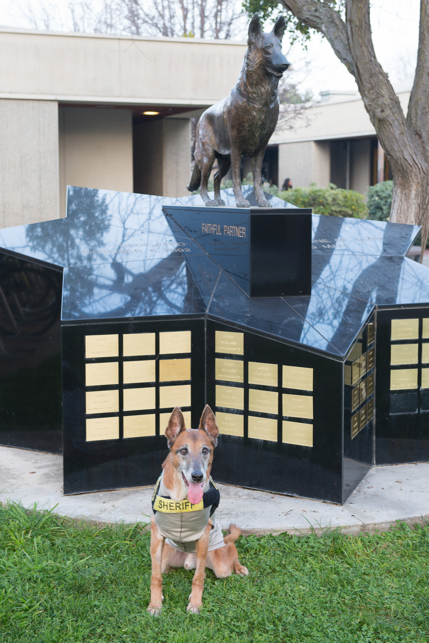
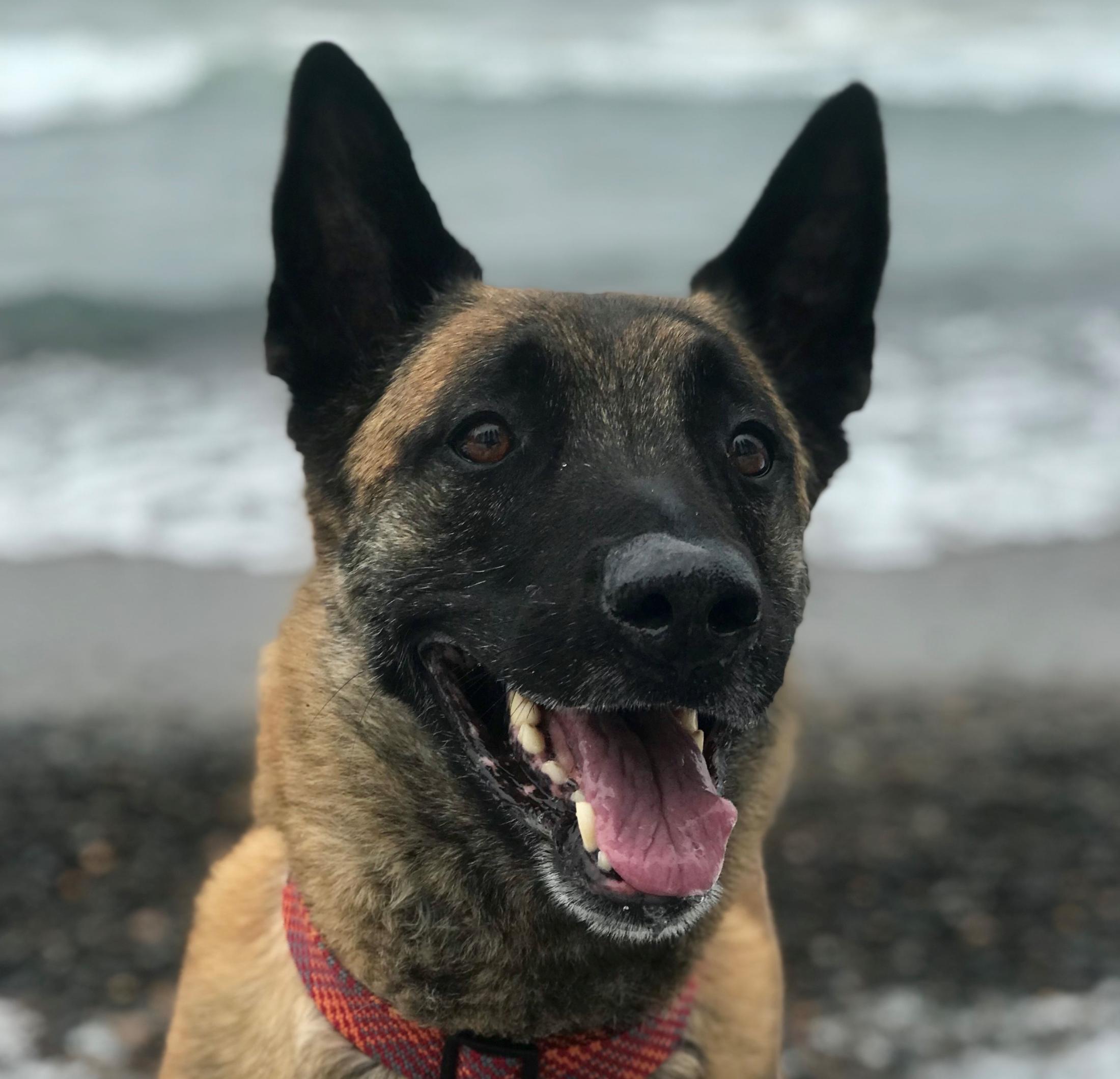
Prostate EmbolizationKopper at the Faithful Partner statue on the UC Davis campusProstate embolization has become a major advancement in the treatment of prostate cancer in animals and was developed for veterinary patients by Dr. Culp at UC Davis. To perform an embolization, a material is injected into the blood vessels supplying the tumor, which causes a blockage of the vessels, thereby cutting off the blood supply and accompanying nutrients to the tumor. The size of the prostate and tumor decrease as cells die from lack of blood supply. Embolization has now become the major local disease therapy for prostate cancer in dogs at UC Davis. Thanks to Dr. Culp’s success with the procedure, many oncologists are considering this as a first-line therapy for prostate cancer and are sending cases to UC Davis. AbelMuch of this work started in 2015 with a dog named Kopper, a 14-year-old Belgian Malinois from Tennessee, who was a retired K-9 officer with the Blount County (TN) Sheriff's Department. Since then, Dr. Culp has performed the embolization procedure in nearly 50 dogs, including Abel, a 10-year-old Dutch shepherd. To obtain a better scope of Abel’s disease and to help plan the surgery, Dr. Culp worked with the hospital’s Diagnostic Imaging Service to perform a CT scan. The results confirmed a mass suspected to be prostatic carcinoma. During the procedure, the blood supply to Abel’s prostate was accessed through his carotid artery. Utilizing fluoroscopy, Dr. Culp was able to navigate to the prostatic arteries where he introduced microscopic beads through a catheter to block the blood supply to the prostate, eliminating the vast majority of the blood supply to the tumor.
Liver Embolization Milo, a 12-year-old male Pekingese, was found to have a large tumor on his liver. Since surgical removal of the tumor was not a viable option and systemic chemotherapy is not generally considered majorly effective for Milo’s disease, Dr. Culp utilized IR to perform a transarterial chemoembolization of the tumor, whereby a catheter was guided through his femoral artery to the tumor’s main blood supply. Once there, beads impregnated with a chemotherapy drug were inserted to cut off the blood supply to the tumor. Over time, this caused a decrease in the size of the tumor and improvement in Milo’s overall status.
Unique Cancer Surgeries and Minimally Invasive Options
Faculty Lead: Dr. Michele Steffey,
Soft Tissue Surgery Service
With new surgical treatments for cancer and minimally invasive options to treat other diseases and injuries, care at UC Davis is being sought after by clients from thousands of miles away.
Learn More
Dr. Michele Steffey, a professor in the Small Animal Soft Tissue Surgery Service, is bringing hope to pet owners with unique procedures. With new surgical treatments for cancer and minimally invasive options to treat other diseases and injuries, her work is being sought after by clients from thousands of miles away.
Thermal Ablation to Treat Cancer: Thermal ablation is a process of killing tumor cells by either heating them (microwave or radiofrequency ablation) or freezing them (cryoablation). Dr. Steffey has pioneered the use of cryoablation to treat several cancers, including adrenal tumors (pheochromocytoma), nasal tumors (adenocarcinoma), and bone cancer (osteosarcoma). Her work has shown cryoablation to be an effective alternative treatment for operable and inoperable deep tissue cancers in veterinary patients.
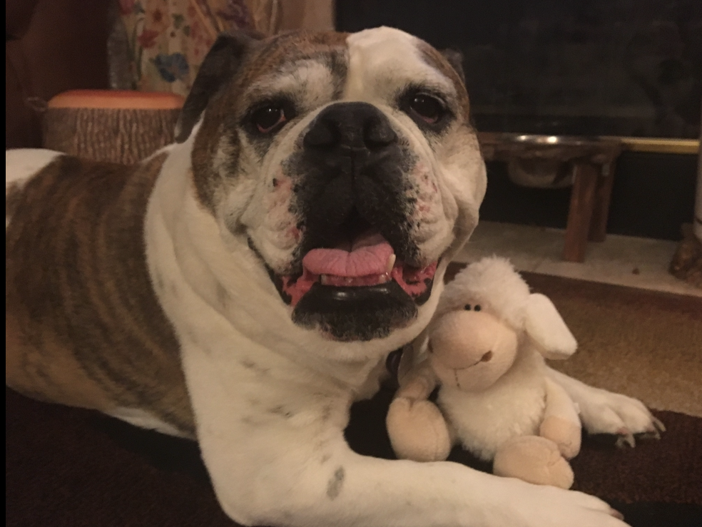
Adrenal Tumors ZoeyDr. Steffey’s novel approach to treating adrenal tumors with cryoablation has given Zoey a new lease on life. The 11-year-old female English bulldog was producing excess adrenaline because of a tumor on her adrenal gland. The excess adrenaline was being randomly secreted into her system and causing heart disturbances. Radiation therapy was successful in stopping the growth of the tumor, but didn’t stop the excess hormone secretion, so Dr. Steffey sought a new path to “turn off” the tumor. Utilizing IR with ultrasound and CT guidance to get the cryoablation probes placed, Dr. Steffey—in collaboration with the Diagnostic Imaging Service and the Anesthesia/Critical Patient Care Service—was able to safely and minimally invasively kill the active tumor cells by freezing the residual tumor tissue. This is the first reported case anywhere in veterinary medicine of treating an adrenal tumor with cryoablation.
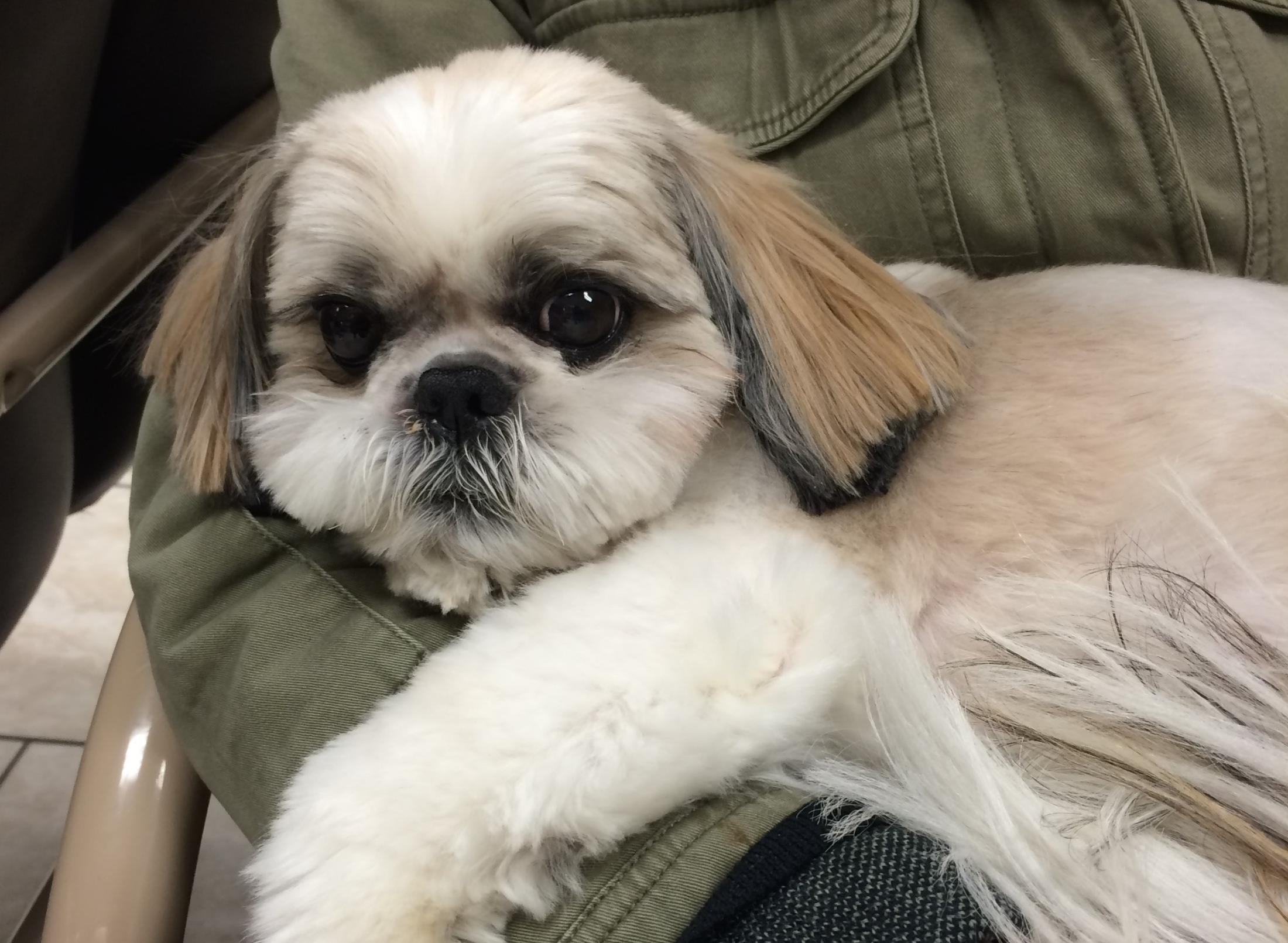
Nasal Tumors JackIn 2013, Dr. Steffey started a clinical trial to test the efficacy of treating canine nasal tumors with cryoablation. After publicizing the success of an early case, dog owners nationwide sought to enroll in the trial. Radiation therapy—consisting of 16 treatments over the course of three weeks—is the gold standard of care for nasal tumors in dogs, and data regarding overall survivals for this treatment show a mean survival time in the range of 12-14 months. However, a number of patients of Dr. Steffey’s cryoablation treatments are now cancer free up to five years post treatment. Jack’s family traveled from Ontario, Canada for the treatment. The 7-year-old male Shih Tzu is showing no recurrence of cancer four years later. Maggie, a 9-year-old female black Labrador retriever from Oregon, remains cancer free three years after treatment. UC Davis remains the only veterinary hospital currently utilizing cryoablation to treat nasal tumors.
Bone Cancer While cryoablation has been utilized extensively to treat bone cancer in humans, it is a very new application in veterinary medicine. A major issue to overcome in veterinary medicine that doesn’t exist in human medicine is aftercare. Most osteosarcomas in animals occur in the distal aspect of the limbs. While humans can use crutches or wheelchairs during recovery, dogs don’t have that option – their recovery is limited and challenging. Dr. Steffey has only performed two procedures on dogs, but feels that it has a future as a viable option for certain types of patients. Continued applications will help measure future risk of fracture in limbs with tumors, improve patient selection for options such as palliative radiation therapy, and aid in the development of alternative limb-salvage procedures.
All of Dr. Steffey’s novel approaches to tumor ablation have involved cryoablation thus far, but she very recently also acquired a microwave ablation unit, which will expand treatment opportunities for different tumor types and locations. She looks forward to advancing the surgery service’s capabilities to regularly include microwave ablation applications in the near future.
The UC Davis veterinary hospital places great emphasis on teamwork throughout the many specialty services in the hospital to provide all-inclusive treatment plans under one roof. Through this collaborative approach to patient care, surgeons and internists are able to bring pioneering mindsets to unusual problems being presented by patients.
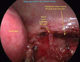
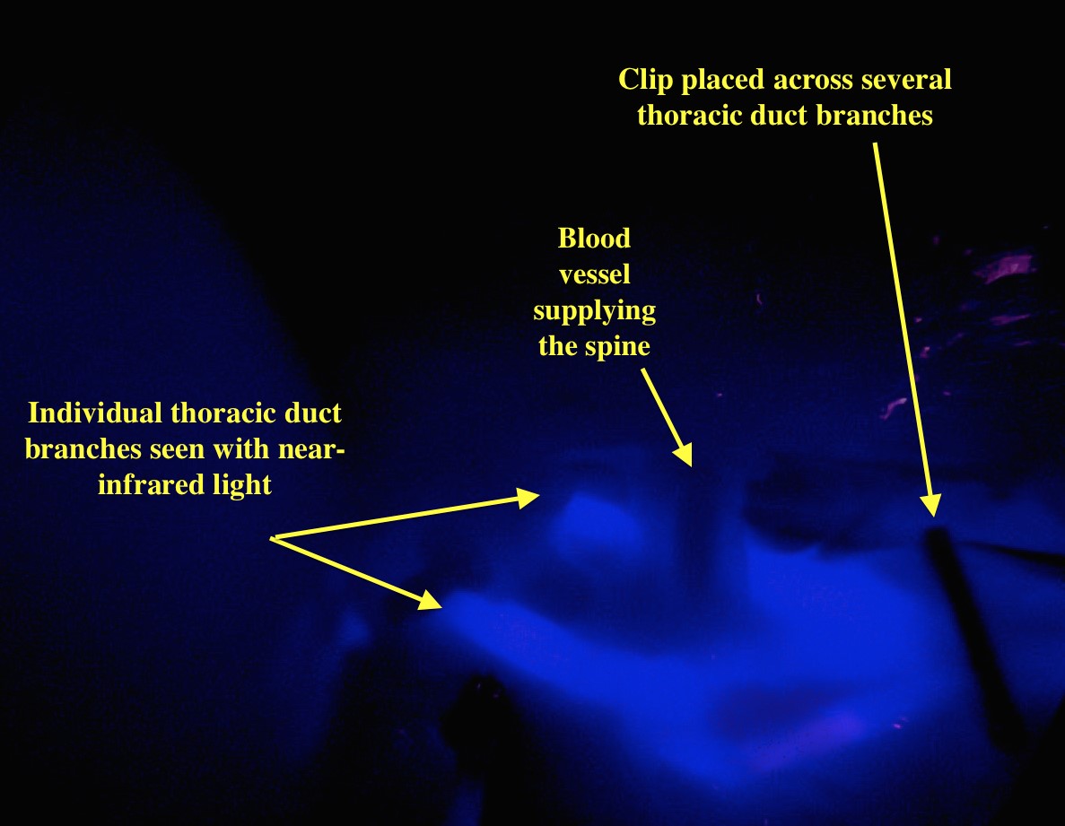
Near Infrared Imaging:Near infrared imaging Pioneered at UC Davis by Dr. Steffey, this exciting new form of surgical imaging uses an injectable tracer pharmaceutical that is detected by a special imaging system at very low doses, with a very high sensitivity, to highlight specific structures in the surgical field that otherwise may be unseen with the naked eye. In the context of cancer surgery, this provides the potential for improvement in the identification of tumors, identification of lymph nodes that should be examined for metastatic disease, and delineation of surgical margins. These improvements increase operative accuracy and decrease surgical time. Unlike many other types of imaging, there is no radiation dose to the patient with near-infrared fluorescent imaging, and this modality can be used in minimally invasive surgery, as well as traditional open procedures. This technique has also been useful for identifying the optimal lymph node for biopsy when assessing for lymphatic metastasis, treatment of chylothorax, and improving dissection for gall bladder surgery. AtlasNear infrared imaging was used to enable thoracoscopic surgery to treat Atlas, a 3-year-old male mastiff, for chylothorax, a build-up of a milky white fluid called chyle (which leaks from the thoracic duct) in his chest. Dr. Phil Mayhew performed surgery that involved a duct ligation to stop the leakage. Dr. Steffey provided the improved imaging to assist in identifying the thoracic ducts, which are very small and often difficult to see at surgery under normal illumination, as they are buried in fat and other tissue. A dye injected into one of Atlas’s lymph nodes traveled into the lymphatic vessels and made them more visible through tissue when seen using a near-infrared view through a scope, and helping the procedure to be completed effectively through a minimally-invasive approach.
Fixing Congenital Conditions: Dr. Steffey has also brought a pioneering mindset and novel approaches to treating congenital defects and injuries in the trachea and esophagus. Working with specialists in the Internal Medicine Service as well as the College of Engineering’s Biomedical Engineering department, Dr. Steffey helped to create artificial rings to treat a congenital defect of the trachea in a dog. Currently, there are no commercially available rings to implant into larger dogs to repair congenital tracheal ring collapse, so Dr. Steffey worked with engineers to help fabricate custom rings for implantation.
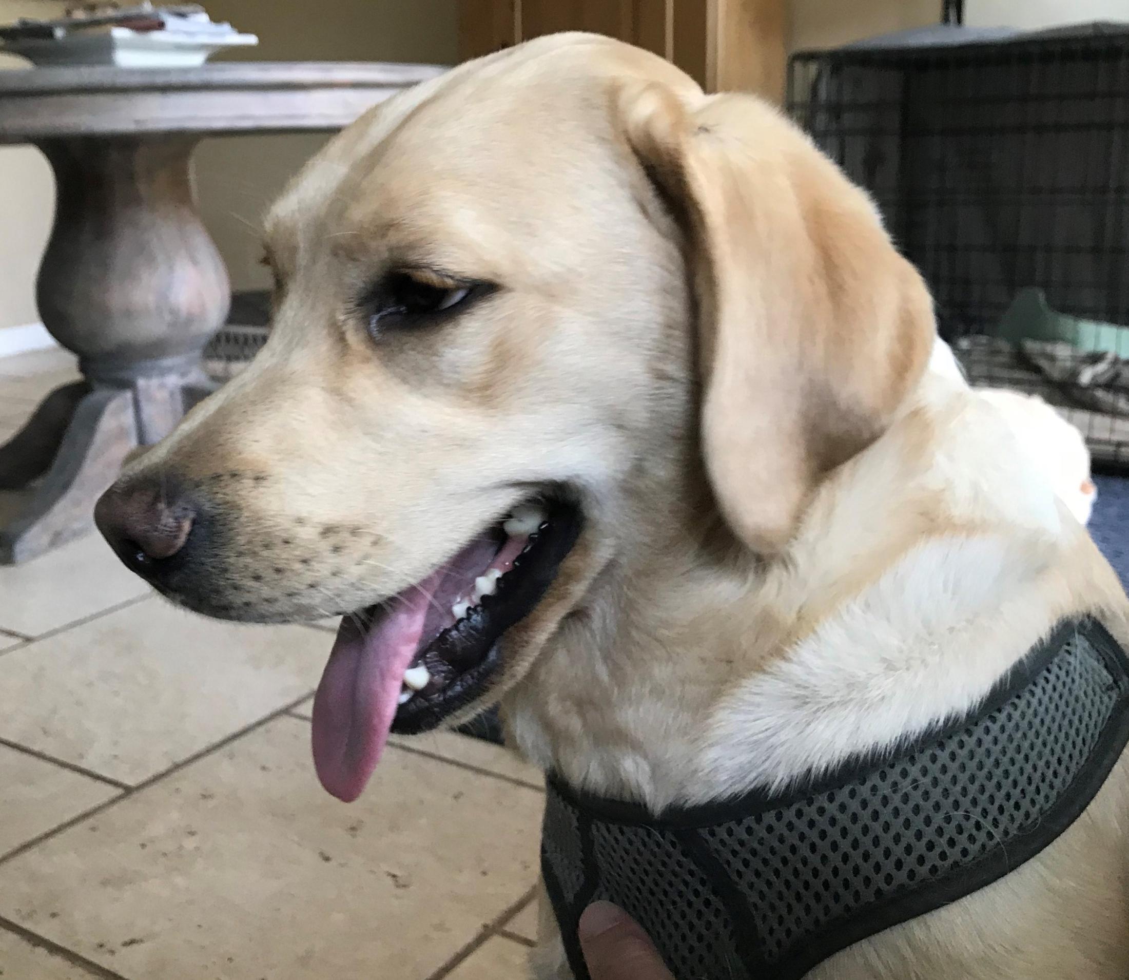
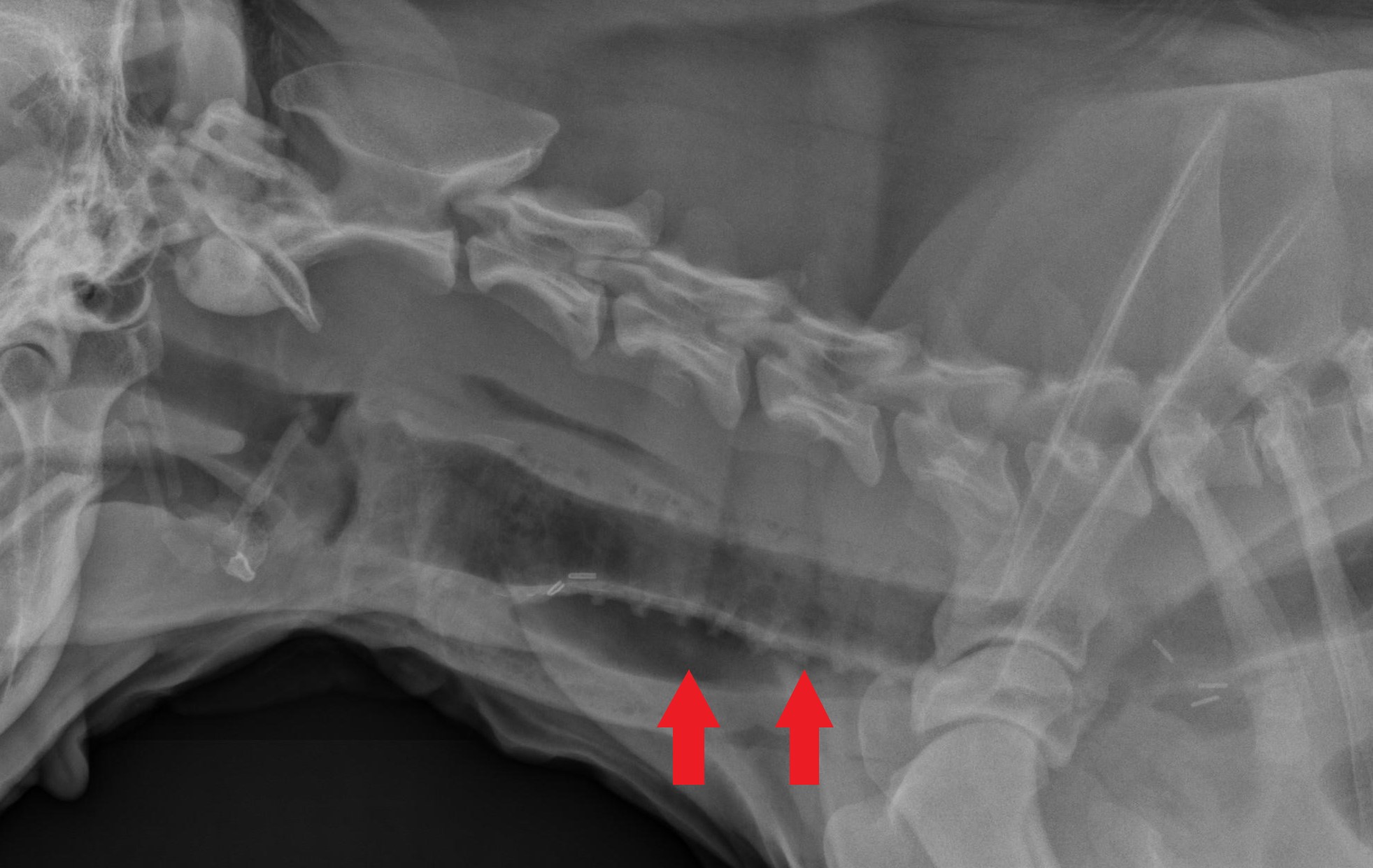
Artificial Trachea RingsBuddyBuddy's x-ray showing the trachea rings in place after surgery Buddy, a 1-year-old male yellow Labrador retriever, has been affected by labored breathing since birth, requiring many visits to and treatment by a number of UC Davis specialists for respiratory distress. To treat this ongoing condition due to a congenital trachea collapse, Dr. Steffey—working in conjunction with Buddy’s primary internal medicine specialist Dr. Sean Hulsebosch, who has dedicated countless hours and concerted effort navigating Buddy’s many issues and directing a team of residents and students to provide his overall care—implanted 15 custom-made rings on his trachea to keep it open. Six months post surgery, Buddy’s breathing is back to normal and his quality of life has greatly improved.
Repairing Esophageal Constrictions Since birth, Sophie, a 1-year-old female collie/retriever mix, had trouble keeping her food down. She would regurgitate food immediately after almost every meal. A CT scan at UC Davis found a persistent right aortic arch (PRAA) – an abnormal development of a major blood vessel in a way that formed a circle around Sophie’s esophagus, trapping her esophagus between the blood vessel and the heart. As a result, the PRAA was preventing her esophagus from expanding to allow food to pass. Dr. Steffey felt that Sophie was a good candidate for a new minimally invasive surgery to correct the developmental defect. She was able to thoracoscopically transect the vessels that were restricting Sophie’s esophagus. After transection, the esophageal constriction was visibly relieved almost immediately. Since the surgery was performed without having to open Sophie’s chest, she recovered quicker and with much less pain. Her owners report that the regurgitations are no longer a regular occurrence.
Maxillofacial Repair
Faculty Leads: Drs. Frank Verstraete and Boaz Arzi, Dentistry and Oral Surgery Service
Over the past decade, the UC Davis Dentistry and Oral Surgery Service team has transformed veterinary oral and maxillofacial surgery.
Learn More
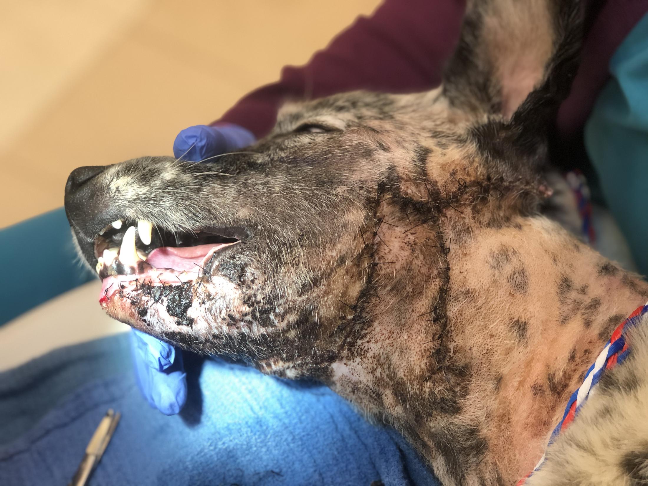
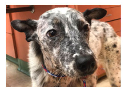
Over the years, Drs. Frank Verstraete and Boaz Arzi of the Dentistry and Oral Surgery Service (DOSS) have transformed veterinary oral and maxillofacial surgery. They have literally written the book on the subject (Oral and Maxillofacial Surgery in Dogs and Cats, 2nd edition now available), and their services are as sought after as any procedures in veterinary medicine. After gaining worldwide acclaim in 2013 for saving Filipino hero dog Kabang (whose snout was torn from her face), the team’s innovations have only expanded. They are pioneering the regrowth of jawbones and have discovered a successful stem cell treatment for a debilitating oral disease that affects one in seven cats. Their latest success involves a massive facial injury that required an innovative skin transformation. Mila after surgery with extensive stitches on her neckMila, a 7-year-old female Australian cattle dog, got into a tussle with another dog through a fence. With her snout extended through the fence, the other dog latched on, leaving Mila with no means to defend herself. With Mila stuck in the compromising position, the other dog ripped the flesh under her chin down her neck.
After being brought to the Emergency Room, where she was assessed and stabilized, Mila was transferred to DOSS for the expert care of the maxillofacial surgeons. To most effectively treat Mila’s injury, the DOSS team needed to perform a cone beam CT scan to determine the extent of the damage. The scan ruled out a mandibular fracture or any other internal injuries.
The team moved to surgery to repair the wound to her chin. The wound was cleaned, and the surgeons were able to reattach the torn skin. However, there was risk of necrosis of this repaired area, and Mila’s owner was warned a second surgery may be needed if the repair was not complete. Mila healed well after several weeks of recovery.If a second surgery was necessary, DOSS planned to transpose skin from Mila’s face and neck to cover the wound. Thankfully, a dog’s skin is elastic and movable in certain areas close to that region. Due to this, Mila’s skin would be able to be repositioned to cover the wound on her chin. Unfortunately, that is what occurred, but a second surgery successfully rotated a flap of skin from the side of her face to cover the exposed tissue on her chin.
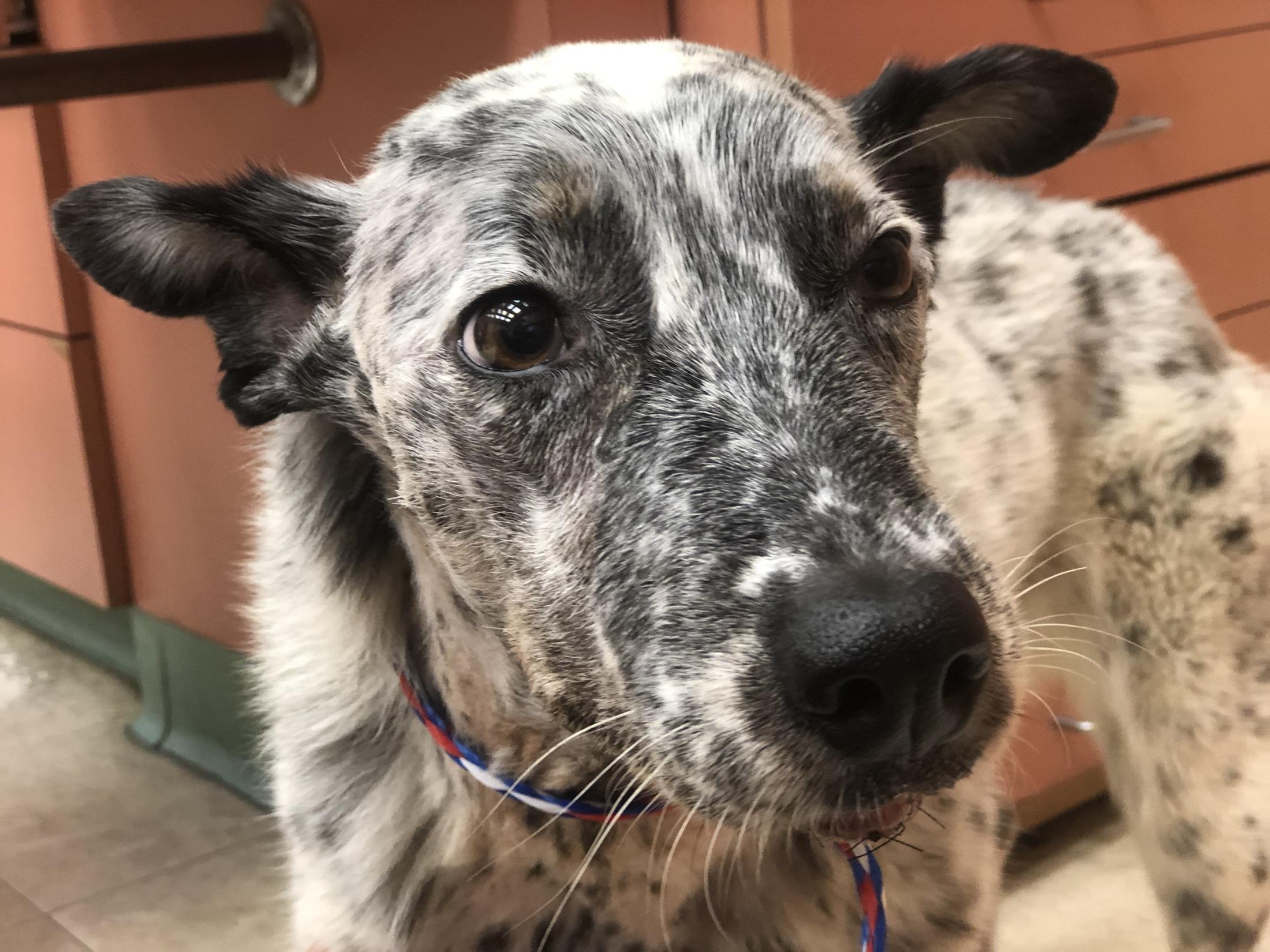
Several recheck appointments post facial reconstruction showed marked improvement as Mila’s wounds healed. She is back to eating and drinking normally and is being given more freedom to play and exercise.
 PET Scanning
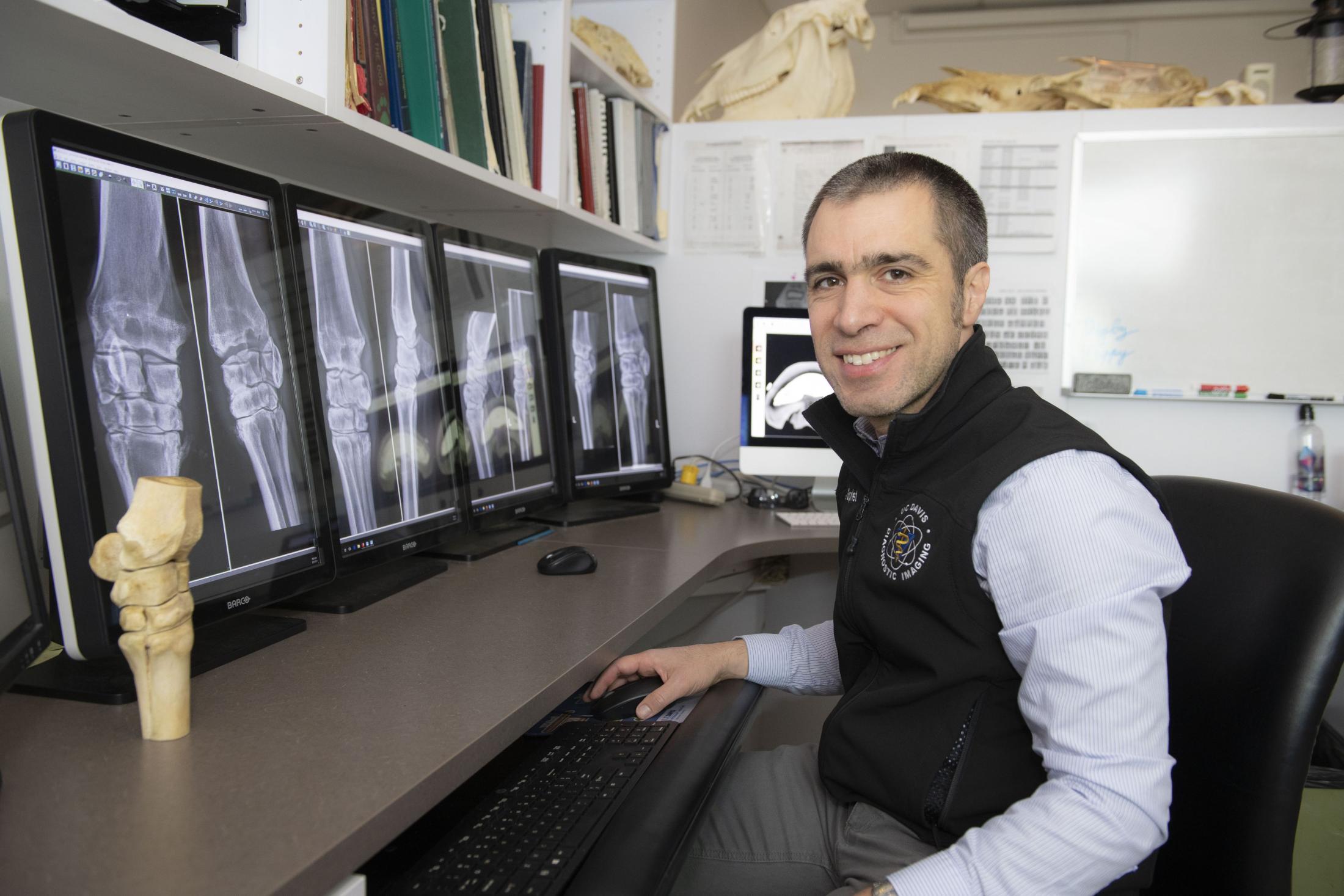
 Data used for both research and clinical applications has shown PET to be particularly useful for detecting bone lesions that are not recognized using other imaging techniques, and to distinguish between active and inactive injuries in bone or soft tissue.
 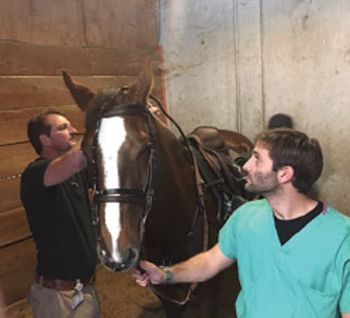 Videoendoscopy to Diagnose Upper Respiratory Disorders
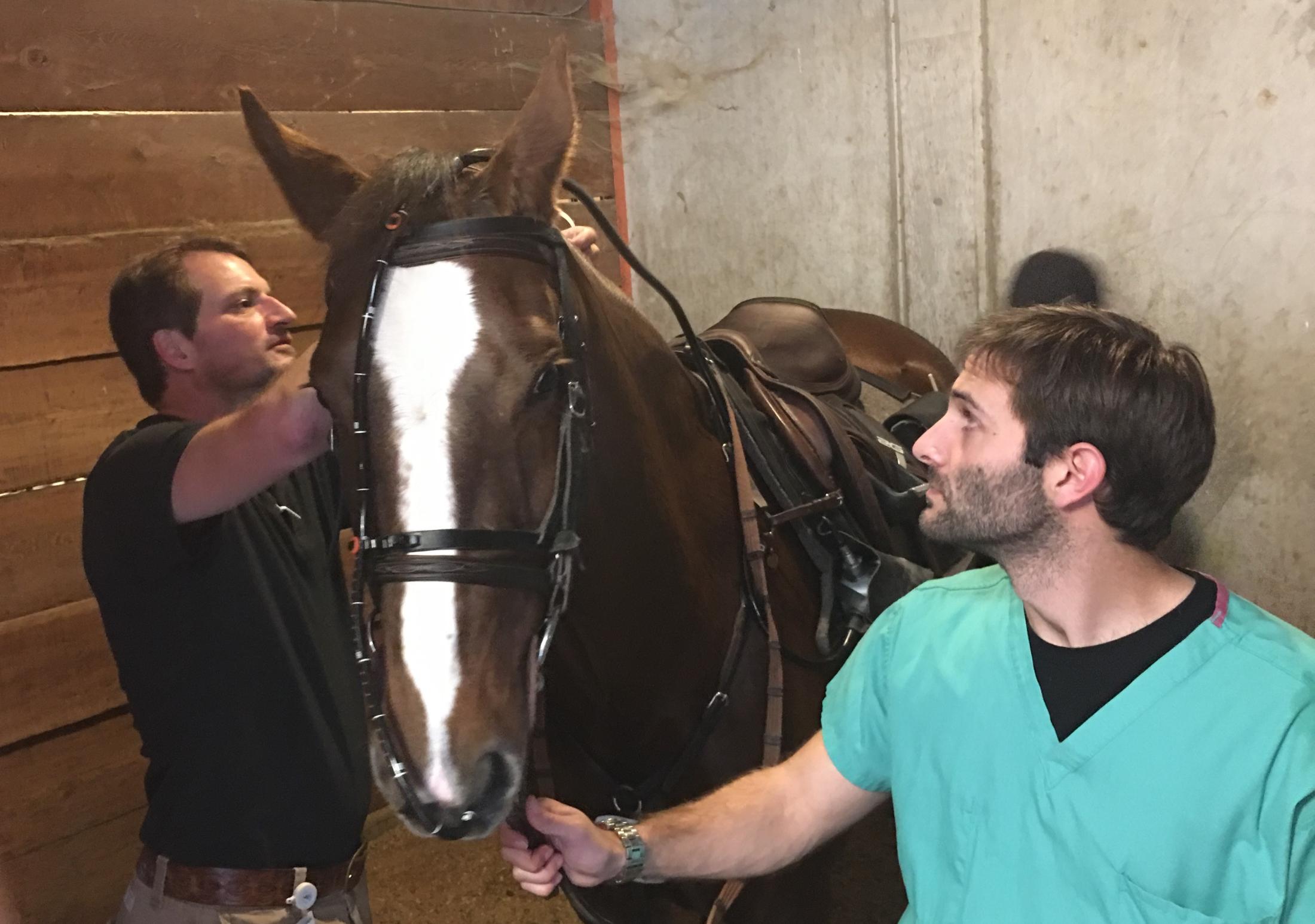 Dr. Scott Katzman, chief of the Equine Lameness and Surgery Service, is utilizing overground videoendoscopy to diagnose dynamic upper respiratory collapse in horses. A malleable endoscope is inserted into the horse’s nasal passage and advanced into the nasopharynx to observe laryngeal movements during ridden exercise. The examination is recorded and transmitted to a small monitor for viewing in real time. With this technology, veterinarians can diagnose causes for upper respiratory collapse not observed during resting endoscopy.
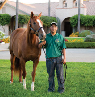
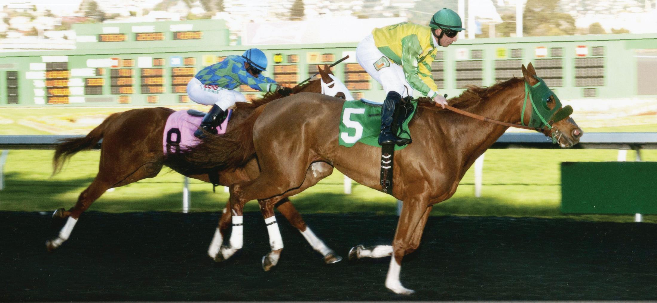 In 2017, racehorse Irish Streetsinger returned to racing following enrollment in Dr. Galuppo’s stem cell trial for an injury that sidelined her in 2016. By early 2018, she ran seven races with one victory at Golden Gate Fields.
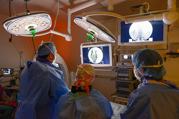
 Maxillofacial Repair