Discoveries
New Insights into Toxoplasma
Toxoplasma gondii is a common parasite hosted by wild and domestic cats and shed in their feces. Although healthy humans rarely experience symptoms, toxoplasmosis can cause miscarriages and neurological disease.
A recent analysis presented by doctoral student Sophie Zhu and colleagues in the journal PLOS One suggests that wild, stray, and feral cats living in areas with higher human population density tend to release—or “shed”—a greater amount of the parasite. The study also draws links between environmental temperature variation and parasite shedding.
While the findings do not establish any causal relationships, they do suggest that rising human population density and temperature fluctuations may create environmental conditions that exacerbate the spread of T. gondii.
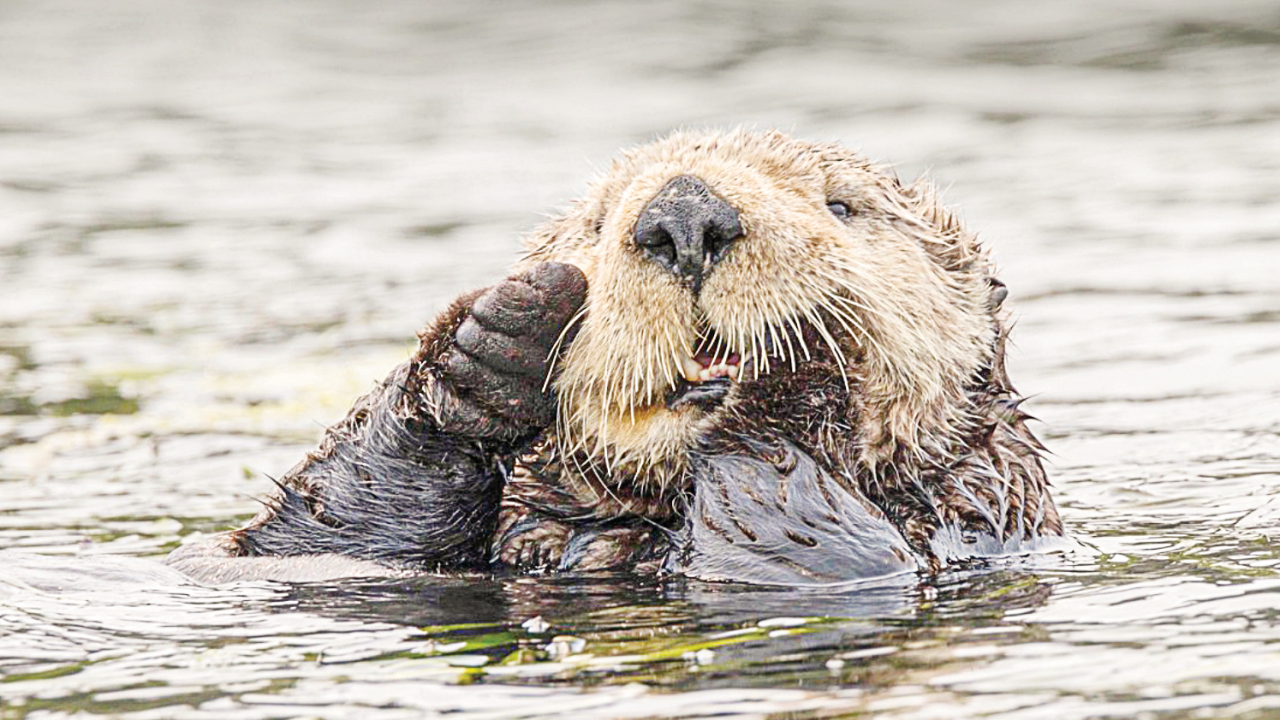
Sea otters are especially vulnerable to Toxoplasma infection because they live near the shoreline where they may be exposed to the parasite’s eggs in rainwater runoff, and they eat marine invertebrates that can concentrate the parasites.
Although toxoplasmosis is common in sea otters, a recent study identified a rare strain of the parasite, previously unreported in aquatic animals, as responsible for the deaths of four sea otters that stranded in California. This rare strain is likely a recent arrival to the California coast. Scientists warn that it could pose a public health risk if it contaminates the environment and the marine food chain.

Steps Toward a Salmonella Vaccine
Salmonella infections cause a million deaths worldwide each year. Improved vaccines for both typhoid fever and non-typhoidal Salmonella disease are urgently needed. New work published in Proceedings of the National Academy of Sciences shows how memory T cells, crucial for a vaccine that induces a powerful immune response, can be recruited into the liver in a mouse model.
“Understanding the immunology is key to developing a better vaccine,” said Professor Stephen McSorley, Department of Anatomy, Physiology and Cell Biology and senior author on the paper.
A type of immune cell known as “tissue-resident memory cells” appears to be key to Salmonella immunity in mice. Usually, when a pathogen enters the body, the immune system mounts a response, which includes CD4 T-cells that support other responses such as antibody production by B-cells. Post infection, some of the cells specific to that pathogen remain as memory cells, ready to respond quickly if the same threat returns. In the mouse model of Salmonella infection, those CD4 memory T-cells don’t circulate around the body. They instead stay in the liver as tissue-resident memory cells.
Claire Depew, a graduate student in the McSorley Laboratory, took CD4 T-cells specific for Salmonella and transferred them into mice that had never been infected. The molecules that promote inflammation, especially interleukin-1 and 2, enhanced formation of Salmonella-specific CD4 tissue resident memory cells in the mice. This provides a rapid response that can act quickly against Salmonella infection. These results will help with the design of new vaccines for Salmonella.

Understanding Equine Evolution
Eighteen million years ago, a genetic duplication event coincided with the evolutionary split between horses and their four-toed, forest- browsing ancestors. A recent study in the journal PLOS ONE, led by Dr. Danika Bannasch and Kevin Batcher, a Ph.D. student in her lab, investigated specific genetic duplications in horses, finding evidence of important roles for these elements.
A certain class of genetic variation in animals is caused by an element called LINE-1 (long interspersed nuclear element 1), which is still active and capable of making new copies of itself and sometimes copies of other genes, called retrocopies. The study investigated these extra gene copies in horses and other domestic and wild equids. Of the 437 retrocopies identified, only five were shared between horses and other equids, indicating that the majority of the extra copies formed after the species experienced an evolutionary split.
In particular, a retrocopy of the gene Ligand Dependent Nuclear Receptor Corepressor Like (LCORL) was present in all equids (horses, donkeys, zebras) but absent from other species of odd-toed ungulates alive today, such as rhinoceroses and tapirs. This retrocopy was also duplicated many times within equids, with the majority of LCORL tissue expression in horses and donkeys originating from these retrocopies rather than the parent gene. Bannasch said this role of LCORL was exciting to identify since the gene is also known to have a significant role in body size across mammals.
The original retrocopy, estimated to have occurred 18 million years ago, happened during the period when equids experienced an increase in body size, a reduction in the number of toes, and changes in their teeth. The age of the LCORL retrocopy and the large number of expressed LCORL retrocopies in today’s equids provide evidence of their function in equid evolution.

Advancing Osteosarcoma Research
A groundbreaking material—engineered bone marrow (eBM)—has the potential to improve treatment for osteosarcoma, a malignant bone cancer with low survival rates. A new study involving UC Davis researchers published in the Proceedings of the National Academy of Sciences describes eBM’s potential. This includes helping researchers learn how bone marrow cells affect osteosarcoma growth, testing cancer therapeutics, and potentially personalizing treatment.
Osteosarcoma is the most common primary bone cancer in children and adolescents, usually affecting children under age 13. Survival rates are low: less than a 25% 5-year survival rate for children with metastatic cancer. It is also the primary bone tumor in dogs, often requiring limb amputation.
Researchers usually study osteosarcoma in flat, artificial cultures that fail to mimic the tumor environment, or mouse models with many variables that scientists can’t control. This new material will allow researchers to better study how these tumor cells grow
and respond to drugs, explained Kent Leach, professor of Orthopedic Surgery and Biomedical Engineering at UC Davis and the corresponding author on the paper.
This work is very exciting because it lays the foundation for a technology that could be used to help veterinary and human patients alike. By providing a realistic bone marrow niche for study in the traditional lab setting, it opens doors for new discoveries.”
—Katherine Griffin, a study coauthor and dual DVM/Ph.D. candidate under Leach’s mentorship through the Veterinary Scientist Training Program at the UC Davis School of Veterinary Medicine. “