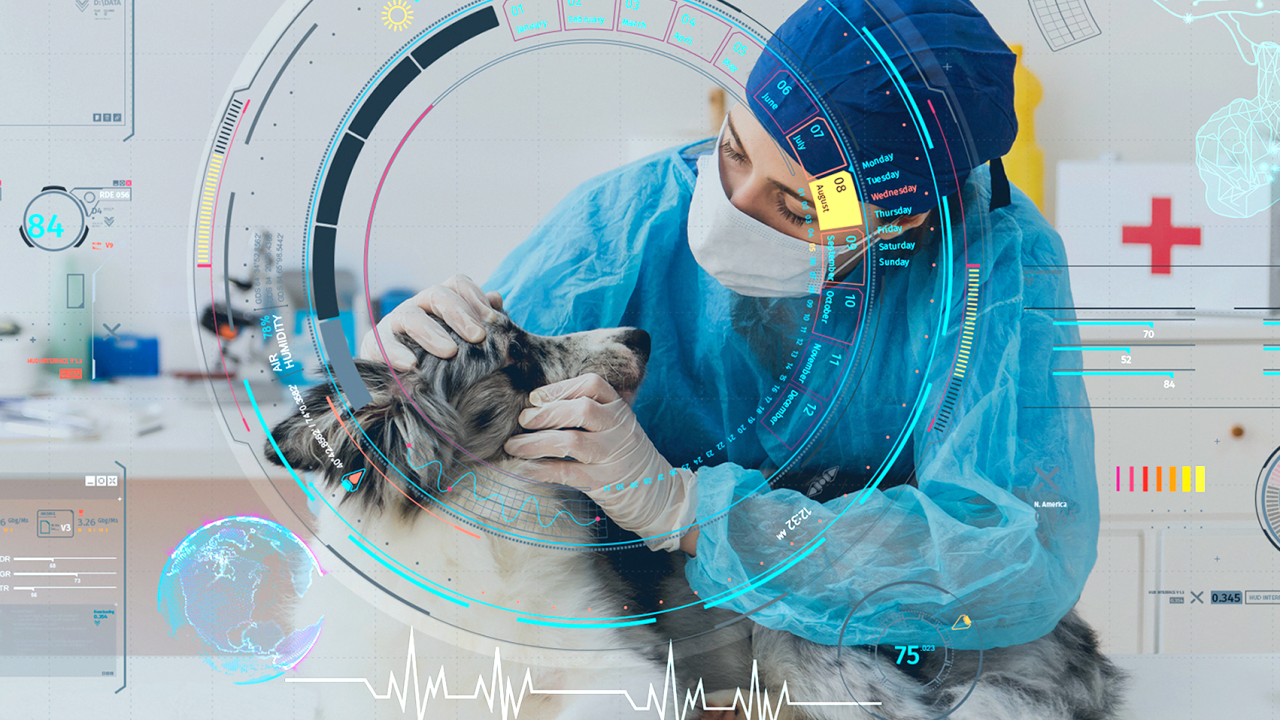
Top Clinical/Research Advancements Shaping the Future of Veterinary Medicine
In our spring issue, we brought you some of the top clinical and research accomplishments from the past 75 years. Here, we delve into the most current and advanced technologies that will take us into the future.
From new discoveries in imaging modalities to artificial intelligence, innovation now drives the practice of healing animals. UC Davis has always been at the forefront of that progress and continues to forge new ground. As we are building the Veterinary Medical Center, we are setting the gold standard of care while defining translational research and clinical education.

Artificial Intelligence
Veterinarians at UC Davis have developed algorithms using artificial intelligence (AI) to detect two significant diseases in dogs—Addison’s and leptospirosis. For Addison’s disease, they used routine blood work from more than 1,000 dogs to train an AI program to detect complex patterns that indicate the presence of the disease.

Their algorithm for Addison’s disease is more than 99% accurate. Next, the team developed an AI prediction model for leptospirosis, which can cause kidney failure (requiring dialysis), liver disease, and severe bleeding into the lungs. When recognized early, the disease can be treated effectively in 90% of dogs. Blood samples from more than 400 dogs, which included only nine that were positive for leptospirosis, were used to train an AI program. When completed, the program achieved 100% sensitivity, correctly identifying all nine samples. Quicker detection through AI will allow veterinarians and pet owners to make critical decisions in a timely manner. These breakthroughs hold tremendous potential for the development of future AI tools to optimize detection of other veterinary diseases. Read more about the uses of AI in veterinary medicine in our feature story.

Advancing Equine PET Scan Technology
In 2015, the world’s first equine positron emission tomography (PET) scan was completed at UC Davis on the leg of an anesthetized horse. Radiologists, working with private industry, continued to advance this new technology, and in 2019 created the first standing PET scanner that only requires a horse to be mildly sedated.

After many prototype iterations, the current MILE-PET® scanner was developed. Research has confirmed the value of PET, especially in the horse racing industry, and the technology has proven superior to bone scans in assessing the racehorse fetlock. PET also demonstrated its ability to monitor injuries over time, predict the amount of time needed to heal, and help prevent catastrophic breakdown injuries. Thanks to UC Davis’ success in pioneering PET, scanners are now in place at some of the most renowned racetracks in the world, including Santa Anita Park and Churchill Downs, as well as leading equine hospitals such as Kentucky’s Rood & Riddle Equine Hospital (founded by UC Davis alumnus Dr. Bill Rood) and the World Equestrian Center Hospital in Florida.

Expanding Orthopedic Surgery
Over the past five years, Orthopedic Surgery Services faculty have taken their program to new heights. With great demand for their services, the school recently opened the Center for Advanced Veterinary Surgery (read the story in this issue). As some of the foremost authorities on orthopedic surgeries, their translational research is setting the standard of care for their specialty.

One of the most sought-after surgeries is a total hip replacement, now able to be performed using custom-fitted, 3D-printed titanium implants. One of the first UC Davis patients to receive this new type of hip implant was Dexter, a German shorthaired pointer, who had been hit by a car. Utilizing exact measurements of Dexter’s anatomy from a CT scan, a new hip was crafted, consisting of a cup affixed to his pelvis using two cortical screws and two locking screws, and a custom stem implanted in the top of his femur (see Dexter's case study). With many breeds predisposed to hip disorders, these surgeries hold great promise for healthier extended lives.

Revolutionizing Soft Tissue Surgeries
The use of real-time imaging modalities to perform minimally invasive procedures—known as interventional radiology (IR)—is revolutionizing surgeries for companion animals. The Soft Tissue Surgery Service is a world leader in this field and has created many therapeutic breakthroughs. Zeke, a 14-year-old beagle/cocker spaniel mix, traveled from Colorado for liver cancer treatment. His owners chose to enroll him in a clinical trial assessing a minimally invasive method of eliminating a tumor’s blood supply; additionally, this novel therapy involved delivering chemotherapy directly to the tumor. Once the blood supply to Zeke’s tumor was mapped during the procedure, the tumor was accessed through minimally invasive fluoroscopic guidance (real time “X-rays”). Catheters placed into the femoral artery were guided by imaging to the tumor. Embolic beads were injected, causing a blockage of the vessels that directly fed the tumor, thereby cutting off its blood supply and accompanying nutrients. The goal of the procedure is to shrink the tumor, prevent further growth, and improve Zeke’s comfort and quality of life. Additionally, the use of minimally invasive surgical and IR techniques is utilized for sentinel lymph node mapping and biopsy procedures, thermal ablation of cancer, the use of intraoperative near-infrared imaging, laparoscopic and thoracoscopic diagnostic and treatment options, and many other ways of improving animal health.

Collaborative Care
The biggest benefit to care at UC Davis is the collaborative nature of the hospital. Faculty from multiple specialties routinely work together on cases. Advancements in surgery, diagnostic imaging, oncology, critical care, and more are a direct result of this. It took a significant collaboration recently to save Davis, a 3-day-old Thoroughbred foal unable to stand.

Initial tests showed evidence of sepsis in his joints and bones, a dangerous inflammatory reaction to a widespread infection. He was hospitalized for three weeks, receiving aggressive antibiotic and supportive therapies including a plasma transfusion, as well as arthroscopy and flushing of his infected joints. Long-term antibiotics were continued, and he had an excellent outcome. His care team included board-certified specialists and residents from the Equine Field, Internal Medicine, Medical Emergency, Critical Care, Neonatology, Diagnostic Imaging, Surgery, and Anesthesia Services, as well as support from the Transfusion Medicine Laboratory, Clinical Diagnostic Laboratory, Pharmacy, and dozens of technicians and students. This collaborative group worked tirelessly around the clock to bring Davis to health. Very few equine hospitals have the extensive team in place to handle all aspects of a case like this in-house. The level of equine care at UC Davis looks to scale even higher with the creation of the Equine Performance and Rehabilitation Center in the coming years.

Advancing Equine Reproduction
The UC Davis Veterinary Assisted Reproduction Laboratory is focused on understanding the complex nature of gamete physiology and embryo development in the horse and other animal species. One of the most exciting advancements by the laboratory is the development of an intracytoplasmic sperm injection (ICSI) program. The technology required for this type of equine reproduction is typically not available from veterinary practices. Unlike the normal process of in vitro fertilization (which generally does not work with horses), ICSI involves injecting a single sperm into an egg extracted from a mare.

The embryo then develops in a laboratory for a week before being implanted in the mare. The process includes aspiration of immature or mature eggs using ultrasound guidance; in vitro culture of the eggs; micromanipulation and microinjection of eggs with a single selected sperm; and laboratory embryo culture, freezing, and transfer to synchronized recipient mares. Recently, Lady Laine was born thanks to ICSI technology, and she is thriving into a healthy filly.

Expanding Emergency Care
Admittances to the UC Davis veterinary hospital’s emergency room (ER) have more than doubled since immediately before the pandemic. Historically, the ER caseload has increased tenfold since 2013, seeing an average of more than 900 cases per month in 2022, with some months seeing more than 1,200 patients. To help meet demand, the hospital opened a new ER and intensive care unit (ICU) totaling approximately 1,600 square feet, nearly doubling its previous size. Funded entirely by donations, the new facility optimizes patient care and expands opportunities for students and emergency specialists.

Recent patients to the new ER included a cat with renal failure after a toxic ingestion, a dog with intervertebral disc disease that ultimately needed neurosurgery, and a sloth named Suzy from the SeaQuest Aquarium who was not responding well to her diet, losing weight, and in poor overall health as a result. After several days of hospitalization, supportive care, and a new diet, Suzy recovered from her illness and regained muscle mass and body weight.

Breakthroughs in Cancer Treatments
Novel cancer treatment breakthroughs are continually being discovered thanks to new technologies and a robust clinical trials program. The recent acquisition of an electrochemotherapy device to treat tumors on or just under the skin is proving to be a valuable alternative method of care.
The treatments involve administering chemotherapy drugs intravenously then sending electronic pulses into the tumor area. The electric current creates pores in the tumor’s cells, allowing a higher percentage of the chemotherapy agent to enter the directed areas, killing the cells and preventing cell division. Other innovative cancer treatments involve immunotherapy—activating the body’s natural killer cells to fight off the growth and spread of cancer. Several clinical trials are investigating the concept, including the use of interleukin-15, a protein inhaled by patients. Tyson, a 10-year-old pit bull terrier mix, saw tremendous results from the trial, which helped stimulate his immune system defenses against his cancer. Tyson is helping bring attention to the science of comparative oncology, which is creating a unique partnership between UC Davis Veterinary Medicine’s Center for Companion Animal Health and UC Davis Health’s Comprehensive Cancer Center, as the two research centers combine forces to fight cancer in both animals and people.


Advanced Ophthalmic Treatments
A cherished member of the UC Davis family was the first canine patient to benefit from new equipment in the Ophthalmology Service. Pint, the retired tee retriever at UC Davis football games, is predisposed to the growth of benign tumors on his eyelids.

Thanks to a new diode laser, ophthalmologists removed Pint’s tumors with an outpatient procedure, and he is recovering well. For most procedures, a dye is injected into the surgical site prior to use. This dye holds therapeutic properties that are activated by the laser in response to a specific wavelength. As in Pint’s procedure, the surgery site cannot be closed with sutures, but the use of the laser is superior to an alternative cryoablation procedure by creating an immediate scab that protects the area from infection, and results in less post-surgical swelling. Most procedures can be accomplished with only sedation, and the technology can be used to treat cancer in and around the eye, as well as many other ocular diseases including glaucoma, iris cysts, distichiasis (growth of extra eyelashes), and laser retina reattachment. Its use in veterinary medicine is scarce, with few hospitals offering this technology.

Elite Level Cardiology Care
With decreased heart function, bulldog Snoopy was at risk of heart failure. He was previously diagnosed with a leading canine congenital defect—severe pulmonary valve stenosis. A balloon valvuloplasty procedure to correct the improper flow of blood out of his heart had failed (not unusual for his breed). An extremely rare alternative is stenting the valve, a procedure now
performed at UC Davis. Less than 10 veterinary teaching hospitals perform this minimally
invasive procedure that accesses the heart intravenously. Snoopy recovered well, and his heart is showing significant improvement. Another cutting-edge cardiac procedure just beginning at UC Davis, and rarely performed elsewhere, is an electrophysiology program, which allows cardiologists to electrically map the heart and identify the cause of arrhythmias (abnormal heartbeats which can cause sudden death). Prior to this equipment’s arrival, clients needed to seek the procedure on the East Coast or pursue medical therapy alone, which is often not a long-term solution. Instead of trying to control the abnormal rhythms with medications, this procedure identifies the affected cells and scars them with an ablation treatment, eliminating their ability to fire abnormally.
